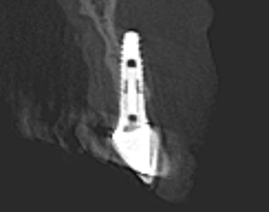
- CBCT scans provide 3D information that increases the safety and accuracy of dental implant placement.
- CBCT Reports written by an Oral & Maxillofacial Radiologist aid in the planning of implants and should accompany each CBCT scan to ensure thorough interpretation of the scan to meet the Standard of Care.
Cone Beam Computed Tomography (CBCT) is an advanced imaging technology that provides three-dimensional views of the teeth, bones, and other structures in the oral cavity and maxillofacial region – these scanned structures should be reviewed by an Oral & Maxillofacial Radiologist who provides the dentist with an official CBCT Report. CBCT has become an essential diagnostic tool in implant dentistry because it provides accurate information about the anatomy and condition of the jawbone, crucial pre-operative information for predictable implant placement.
CBCT scans provide detailed information about the thickness, density, contour, and quality of the bone, as well as the location of vital structures such as nerves, blood vessels, and sinuses. This information is used to plan the optimal placement of dental implants and/or the need for bone grafts, minimizing surgical risks and enhancing long-term success. The use of CBCT for dental implants has several advantages over traditional two-dimensional (2D) imaging techniques such as periapical X-rays.
One of the main advantages of CBCT is that it provides a three-dimensional view of the jawbone, which allows for better visualization and understanding of the anatomy. This helps the dentist to accurately assess the available bone for implant placement and to determine the optimal angle, depth, and position of the implant. CBCT scans also allow for the evaluation of the bone density to some extent, which is important in determining the type of implant to be used and the amount of force that can be applied to the implant.
Another advantage of CBCT is that it reduces the risk of complications during and after implant placement. The use of CBCT scans allows the dentist to identify any potential anatomical challenges such as the presence of nerves, sinuses, or deep concavities in bones that may increase the risk of implant placement. This information helps the dentist to plan and execute the implant placement with precision, reducing the risk of damage to surrounding structures.
To use CBCT for dental implants, the patient is positioned in a special chair or scanner, and the scanner rotates around the head, taking multiple X-ray images. The images are then reconstructed into a three-dimensional model of the jaws, which can be viewed on a computer screen. The dentist can use specialized software to manipulate the images and view them from different angles, allowing for better visualization of the anatomy.
Preoperative CBCT scans are used to assess the patient’s bone quality, density, and quantity, and to plan the optimal placement of the implant. CBCT can also be used during the surgery in the event of any uncertainty to guide the placement of the implant and to ensure that it is positioned correctly. After implant placement, CBCT scans can be used to evaluate the success of the procedure and to monitor the healing process.
CBCT is essential in dental implantology because it provides accurate information about the anatomy and condition of the jawbone, which is crucial for successful implant placement. CBCT scans allow dentists to plan the optimal placement of dental implants with precision, reducing the risk of complications and ensuring long-term success. If you are considering dental implants, ask your dentist about the use of CBCT scans to ensure the best possible outcome.

