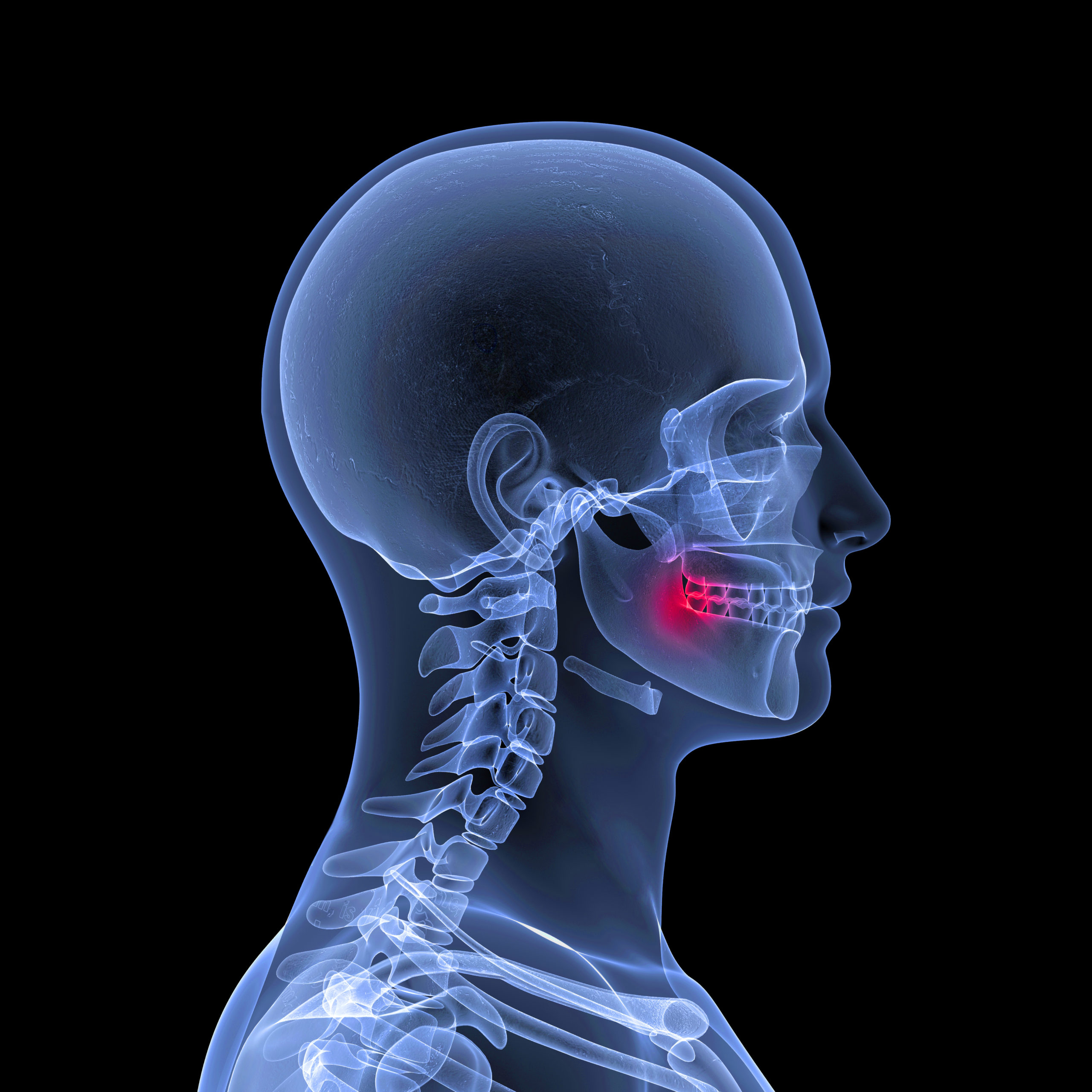
- Cone Beam CT is a type of medical imaging used in dental practice.
- CBCT provides detailed 3D images of a patient’s teeth, jaw, and surrounding structures.
- CBCT Reports can be used to guide procedures such as dental implants, orthodontic treatment, and oral surgery.
- CBCT Reports can also diagnose conditions that affect oral and overall health, such as dental decay, cysts, tumors, and periodontal disease.
- Although CBCT does come with some risks, these risks can be minimized through proper imaging protocols and radiation safety measures.
Cone Beam Computed Tomography, or CBCT for short, is a type of medical radiographic imaging technique that has become increasingly common in dental practices over the past few decades. This technology uses a special type of x-ray machine to capture highly detailed, three-dimensional images of a patient’s teeth, jaw, and surrounding structures. A CBCT Report, typically authored by a specialty-trained Oral & Maxillofacial Radiologist, can provide valuable information that is difficult or impossible to obtain through other diagnostic methods, making it an important tool in the dental office.
So, what exactly can a CBCT Report reveal? For one, it can provide a highly accurate picture of a patient’s dental anatomy, including the position and orientation of each tooth, the shape and size of the jawbones, and the location of important structures such as nerves and blood vessels. This information can be invaluable in planning and executing procedures such as dental implants, orthodontic treatment, and oral surgery, as it allows dentists and oral surgeons to see exactly what they are working with and avoid—or at least be well-prepared to manage—potential complications.
In addition to providing detailed anatomical information, a CBCT Report can also be used to diagnose a wide range of dental and oral health conditions. For example, it can be used to identify the presence of cysts or tumors, apical lesions, and evaluate the extent of bone loss due to periodontal disease. By providing dentists with this detailed diagnostic information, a CBCT Report can help them make more accurate diagnoses and develop more effective treatment plans for their patients. When appropriate, recommendations for management or additional diagnostic procedures are included.
Of course, like any medical imaging technique, CBCT does come with some risks. The main risk associated with CBCT is radiation exposure, as the imaging process involves a small amount of ionizing radiation. However, the amount of radiation used in a CBCT scan is generally much lower than that used in traditional CT scans, and most patients are only exposed to radiation once or twice over the course of their treatment. Additionally, dentists can take steps to minimize radiation exposure, such as by using lower-dose imaging protocols and limiting the number of scans performed.
In conclusion, CBCT is an important tool in the modern dental office, providing dentists and oral surgeons with detailed anatomical information and diagnostic capabilities that are difficult or impossible to obtain through other methods. While there are some risks associated with CBCT, these risks are generally minimal and can be minimized through proper imaging protocols and radiation safety measures.

