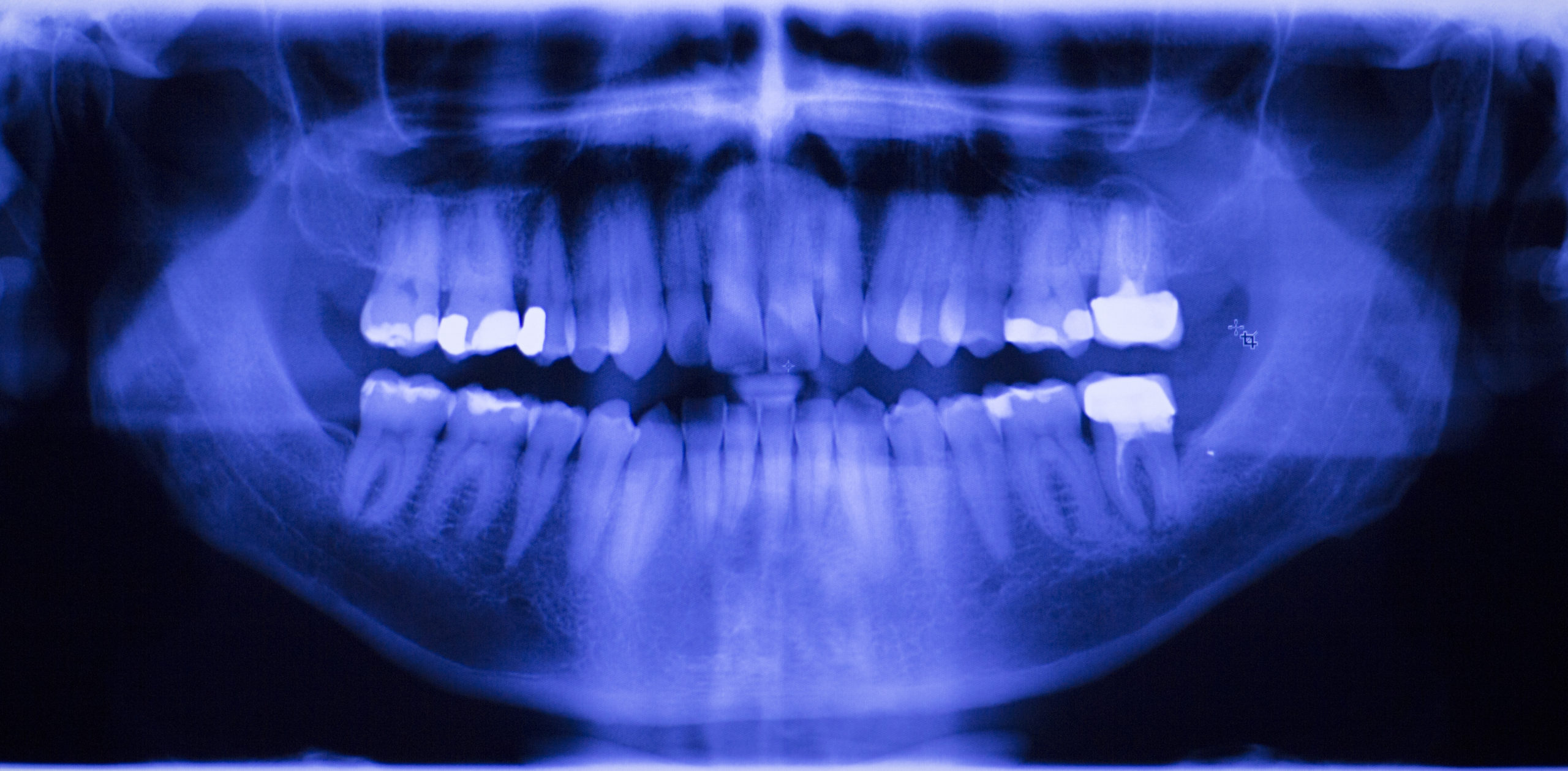
- Radiography is the use of x-rays in dental practice to create images of the teeth, bones, and surrounding tissues.
- Radiographs help dentists identify cavities, bone loss, infections, tumors, and other abnormalities that may not be visible during a clinical examination.
- Radiographs are safe and effective when used appropriately, and they play a critical role in maintaining oral health.
Radiography is an essential tool in dental practice that involves the use of x-rays to create images of the teeth, bones, and surrounding tissues. Although commonly referred to as “x-rays”, these images are technically called radiographs, and they help dentists to diagnose and treat various dental and maxillofacial conditions that may not be visible to the naked eye. Dentists use radiographs for a variety of reasons. They help identify cavities, bone loss, infections, tumors, and other abnormalities that may not be visible during a clinical examination. Radiographs can also help dentists plan and evaluate treatments such as braces, implants, and extractions. Additionally, radiographs can be used to monitor the progression of certain dental conditions over time.
X-ray photons are packets of energy produced in a tube head by heating a tungsten filament—the cathode—causing emission of electrons. Voltage is applied and the electrons race towards the anode—a tungsten disc which, upon certain interactions with the electrons generates the photons. Radiographs are produced by passing x-rays through the body, which are then captured on a detector, either a special type of film or a digital sensor.
The x-rays are absorbed and scattered differently by different tissues in the body, which creates a contrast in the resulting image. For example, teeth and bones appear white or gray on a radiograph, while soft tissues such as gums and cheeks appear darker. Air is the least-absorbing entity in the body, so airways and sinuses appear darkest on radiographs. Enamel is the densest object in the body so appears lightest on the images, although metallic dental fillings and implants appear even more white. Other objects like glasses, hearing aids, and jewelry will also be depicted if not removed, all of which deteriorate image quality due to masking the underlying anatomy and generating excessive artifact.
There are two main types of radiographs used in dental practice: intraoral and extraoral. Intraoral radiographs are taken from inside the mouth and provide detailed images of individual teeth and the surrounding bone. Bitewing and periapical radiographs are the most common intraoral types. Bitewings depict the molar and premolar teeth and include both the maxillary (upper) and mandibular (lower) teeth simultaneously. Bitewings are the best for identifying tooth decay on the tooth surfaces that contact adjacent teeth (interproximal caries) as well as the supporting bone level to assess for the presence or progression of periodontal disease. Periapical radiographs only depict selected maxillary or mandibular teeth in each image and include the entirety of the tooth with its root and supporting bone. A complete mouth series of radiographs would include 20 radiographs—4 bitewings and 16 periapices—or fewer for a patient without a full set of teeth.
Extraoral radiographs are taken from outside the mouth and provide a broader view of the entire jaw and skull. Panoramic radiographs generate a two-dimensional view of the entire maxilla and mandible, allowing a broad view of the teeth, their supporting bone, the temporomandibular joints, and even the maxillary sinuses. Cone beam computed tomography (CBCT) generates a three-dimensional image. The image contents depend on the size of the volume acquired, which itself is dependent upon the machine capabilities and desired resolution. CBCT has granted dentists the ability to locate anatomical structures in advance of clinical intervention, allowing for safer procedures and more predictable outcomes.
Despite their many benefits, some may be concerned about the safety of radiographs. However, modern dental x-ray equipment and techniques are designed to minimize radiation exposure as much as possible. Dentists also take precautions such as using lead aprons and thyroid collars to protect patients from unnecessary exposure to radiation, although the exposure is so minimal, the protective effect of these precautions is mostly psychological for an apprehensive patient, as dental x-rays have not proven to be carcinogenic or harmful.
Radiography is an essential tool in dental practice that helps dentists diagnose and treat various dental conditions. By using x-rays to create images of the teeth, bones, and surrounding tissues, dentists can identify problems that may not be visible during a clinical examination. Radiographs are safe and effective when used appropriately, and they play a critical role in maintaining oral health.

