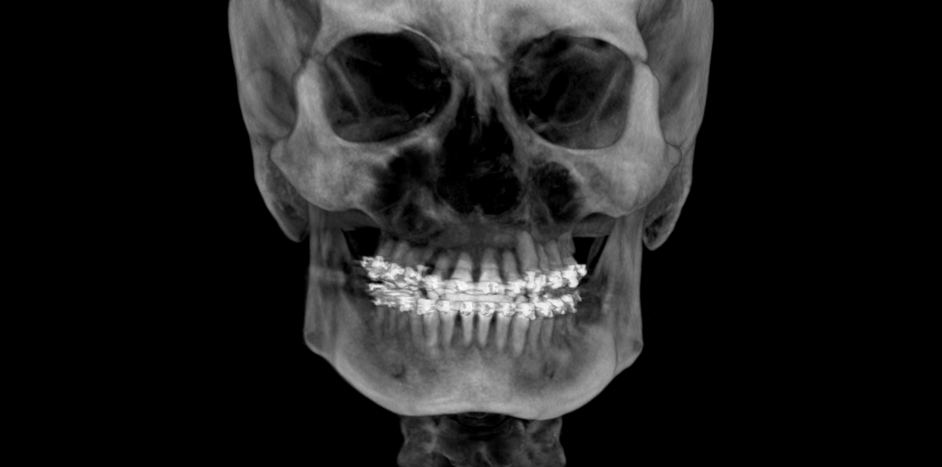
- CBCT generates a 3-D view of the jaws, allowing precise orthodontic diagnosis and predictable treatment planning.
- Judicious use of CBCT before, during, and sometimes after orthodontic treatment will enhance patient outcomes in comprehensive cases.
- The availability and simplicity of CBCT predisposes its misuse.
- CBCT Reports written by Oral & Maxillofacial Radiologists via thorough interpretation of the orthodontic large field of view scans ensure proper documentation of existing conditions and treatment of occult pathology.
Cone-beam computed tomography (CBCT) has become an increasingly popular diagnostic tool in orthodontics due to its ability to provide three-dimensional (3D) images of the craniofacial complex with relatively low radiation doses. This technology has revolutionized orthodontic diagnosis and treatment planning, enabling orthodontists to obtain detailed information on the dental and skeletal structures of the patient, allowing greater accuracy in treatment planning. Several orthodontic indications will be discussed.
Assessment of Dental and Skeletal Structures: CBCT provides a detailed, 3D representation of the craniofacial complex, enabling orthodontists to accurately assess the position, orientation, and size of teeth within the alveolar bone. This information can be used to determine the degree of intervention required for optimal outcomes and estimate treatment length.
Detection of Pathology: CBCT can be used to detect pathology such as cysts, tumors, and dental conditions less detectable with traditional two-dimensional (2D) radiographs – the Standard of Care includes providing a CBCT Report to the level of one written by an Oral & Maxillofacial Radiologist. Orthodontic intervention without acknowledgement of existing conditions is poor patient management and an invitation for litigation.
Airway Analysis: CBCT can provide valuable information on the airway, including its size, shape, and tonsillar classifications. This can be useful in diagnosis of obstructive sleep apnea and other airway-related problems.
Impacted Tooth Evaluation: CBCT is particularly useful in evaluating impacted teeth, as it provides a 3D image of the tooth, its position, and its relation to adjacent structures. This information can be used to plan the appropriate treatment, whether extraction or exposure and orthodontic alignment.
Surgical Planning: CBCT can be used to plan orthodontic and orthognathic surgery, allowing the surgeon to visualize the anatomy of the surgical site and plan the procedure with a high degree of precision.
When using CBCT for orthodontic treatment, it is important to follow the principles of ALADA (as low as diagnostically acceptable), which means minimizing the radiation dose to the patient while still obtaining the necessary diagnostic information. The radiation dose of a CBCT scan is typically higher than that of a traditional 2D radiograph, but it is still considered safe when performed within the guidelines of the American Academy of Oral and Maxillofacial Radiology (AAOMR).
Orthodontists should also be aware of the potential risks associated with ionizing radiation, such as the risk of cancer induction and the potential for thyroid and salivary gland damage. To minimize these risks, the single greatest action the orthodontist can take to prevent excess exposure is avoid unnecessary scans. For example, CBCT for every patient as a screening tool is not appropriate. Additional precautions include the use of lead aprons and thyroid collars.
Biologic risks are only part of the story. Metal is a source of significant radiographic artifact due to its high attenuation. CBCT scans during orthodontic treatment will exhibit artifact due to the metal brackets and wires, an unavoidable fact that must be acknowledged if mid-treatment scans are to be acquired. Details requiring high resolution for interpretation will be very challenging, if not impossible, to detect.
In conclusion, CBCT is a valuable diagnostic tool in orthodontics, providing detailed 3D views of the craniofacial complex that can be used for accurate diagnosis and to enhance treatment planning. Orthodontists should be familiar with the uses and potential risks of CBCT and should integrate the technology appropriately to optimize patient outcomes.

